Understanding
Eye Health
Comprehensive educational resources on eye conditions affecting adults and children.
Eye Health Education
Eye Conditions We Monitor & Manage
From age-related changes to systemic diseases that affect the eyes, our team monitors and manages a wide range of conditions. Select a topic below to learn more.
Cataract
Clouding of the eye's lens. Learn about symptoms, risk factors, and modern surgical treatment.
Learn MoreAdult Eye Conditions
Cataract Symptoms & Treatment
Overview
Cataracts is a clouding of the lens of the eye, resulting in a gradual loss of vision. The lens is the structure just right behind the iris (the colored part of the eye). Most cataract types are related to aging — by age 80 more than half of Americans either have a cataract or have had cataract surgery. Two other common non-age related types include traumatic cataracts and those related to long-term corticosteroid use (like prednisone and cortisone).
How Cataracts Develop
In the healthy state, the transparent lens allows the passage of light through to the retina. When a cataract forms, the cloudy lens does not allow as much light through and vision becomes dull or blurry, and colors lose their brightness. Lights at night may have halos around them.
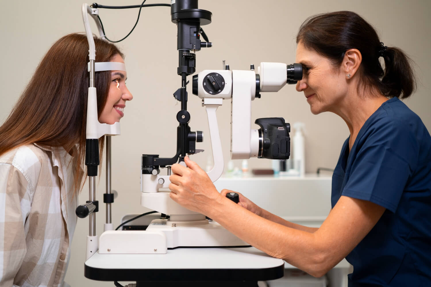
The lens is made mostly of water and protein. As we age, the proteins clump together creating a loss of transparency. The doctor notices the color of the lens becoming a yellow-green. Generally, cataracts cannot be seen with the naked eye — external, surface ocular scars are not cataracts, though many people call them that. A slit-lamp microscope is used to determine the presence or absence of the problem.
Risk Factors
- Age (primarily)
- Certain diseases like diabetes
- Smoking and alcohol use
- Steroid use
- Prolonged exposure to UV radiation in sunlight — UV protection recommended in all eyewear including sunglasses
Treatment
In early stages, prescription changes may improve vision. Surgery is usually required as the condition escalates. Cataract surgery is the most common surgical procedure in the United States, with 90% of patients achieving better vision afterward. The procedure is performed on an outpatient basis — the cataract is removed and an artificial lens implant is inserted.
Post-Operative Care
Eye drops are prescribed for healing and infection prevention. Heavy exercise is discouraged for several weeks following the procedure.
Advanced Techniques
Modern surgeons can calculate implant power to leave patients requiring minimal distance correction. Insurance (usually Medicare) covers a large part of the fees. Patients may pay more for a multifocal implant that provides vision at all ranges.
After-Cataract
Some patients develop clouding of the tissue surrounding the implant months or years later. This is easily fixed by a quick, painless laser procedure.
Key Facts
- Affects 50%+ of Americans by age 80
- 90% success rate with surgery
- Outpatient procedure, same day
- Insurance usually covered (Medicare)
- Modern implants can eliminate the need for distance glasses
Adult Eye Conditions
Diabetes & Your Vision
Overview
Diabetes is a disease in which the body either does not produce enough insulin or does not properly use the insulin it does produce. It affects approximately 17 million people in the United States, and both Type 1 and Type 2 diabetics are at risk for vision loss.
Prevalence
Between 40–45% of Americans with diabetes have some stage of diabetic retinopathy. Each year, 12,000 to 24,000 Americans lose their sight. Diabetic retinopathy is the leading cause of new blindness in U.S. adults.
How Diabetes Affects the Eyes
The blood sugar imbalance can cause weakening of blood vessel walls and seeping of fluid or blood into the retina. As areas swell, tissue can die, causing vision loss. Progression of leakage leads to progressive swelling and further vision loss. Diabetic retinopathy is usually severe before the patient notices any changes.
Annual Exams Are Essential
It is absolutely essential that diabetics have a full eye examination annually. Baseline digital retinal photos are needed after diagnosis, and annually if retinopathy is present. Early detection can prevent or delay serious vision loss.
What We Look For
- Any leaking blood vessels
- Retinal or macular swelling
- Telltale fatty deposits in the retina
- Damaged nerve tissue
- Signs of glaucoma or cataracts, which are more common in diabetics
Treatment
Scatter laser surgery can slow or prevent progression of diabetic retinopathy. New injectable pharmaceuticals are also very effective. The primary goal is to prevent further damage.
The Role of Blood Sugar Control
The potential for eye problems with diabetes can be lessened dramatically with good blood sugar control. Working closely with your physician to manage your diabetes is one of the most important steps you can take to protect your vision.
Warning Signs
- Blurred or fluctuating vision
- Floaters or dark spots
- Partial vision loss
- Difficulty with color vision
- Annual exams are critical for early detection
Adult Eye Conditions
What is Glaucoma?
Overview
Glaucoma is a group of diseases that damage the optic nerve resulting in loss of vision or even blindness, often accompanied by an increase in intra-ocular pressure. It is the second leading cause of blindness worldwide, affecting an estimated 65 million people. Glaucoma is usually painless and often very advanced before the patient notices any changes.
Risk Factors
- Race — up to 8× more common in African Americans
- Age — more common after 60, especially in the Hispanic population
- Family history — 7% more risk if a family member has the disease, ~15% if a sibling
- Steroid users
- History of ocular trauma
- History of hypertension and/or diabetes
- High myopia (nearsightedness)
Four Diagnostic Methods
- Intra-ocular Pressure (IOP) — Not weighted as heavily as before; some patients have normal IOP yet still have glaucoma (normal tension glaucoma).
- Optic Nerve Appearance — The optic nerve looks like a donut where it enters the back of the eye. Glaucoma causes enlarging of the "donut hole" (optic cup).
- Visual Field — Maps each eye's field of view. Typical losses in early glaucoma usually progress from the outside inward.
- The OCT — A laser scan analyzing nerve fiber layer thickness, compared to age- and sex-matched norms.
Types of Glaucoma
Primary Open Angle Glaucoma (POAG) — The most common form. Drainage canals become clogged. Progresses slowly and responds well to medication.
Angle Closure Glaucoma — A medical emergency. Pressure rises quickly, accompanied by ocular pain, nausea, halos, and blurred vision. Requires immediate treatment.
Secondary Glaucomas — Including traumatic, pigmentary, neovascular, and pseudoexfoliative types.
Treatment
Treatment focuses on eye drops and surgery aimed at lowering intra-ocular pressure. Surgery is considered when medication is no longer sufficient to control the disease.
Key Points to Remember
- The disease is painless with no early symptoms
- It is never cured — monitoring never stops
- Treatment is usually effective in halting further vision loss
Did You Know?
- Glaucoma affects 65 million people worldwide
- Often called "the silent thief of sight"
- 7× more common in African Americans
- Regular eye exams are the only way to detect it early
- Treatment halts progression — vision lost cannot be restored
Adult Eye Conditions
Macular Degeneration
Overview
The macula is the area of our retina responsible for central vision. Age-related Macular Degeneration (AMD) is a condition in which the macula is damaged, causing distortion or loss of central vision. It is the most common cause of severe vision loss among people over 65.
Types of AMD
Dry form (Non-exudative) — The most common type. Receptors in the macula slowly break down. Yellowish deposits called "drusen" can be seen during an exam. Vision loss is usually less severe than with wet AMD.
Wet form (Exudative) — Abnormal and fragile vessels grow under the macula. These vessels leak blood and fluid, causing rapid and severe damage. Vision loss is usually devastating and can occur quickly.
Stages
AMD progresses through Early, Intermediate, and Advanced stages. Any wet AMD is classified as "Advanced." The dry form can progress to the wet form over time, making regular monitoring essential.
Nutritional Research (AREDS & AREDS 2)
The landmark AREDS study (National Eye Institute, October 2001) showed that high levels of antioxidants and zinc reduce the risk of advanced AMD by 25–28%. The AREDS 2 study (2006) showed an additional ~12% decreased risk with a refined formula.
High concentrations of these nutrients are not obtainable from a normal diet and are available in supplements such as EyeCaps, Preservision, or OcuVite. Smokers should avoid beta-carotene. Most doctors recommend the AREDS 2 formulation.
Lutein
Lutein has been linked to a reduced risk for AMD. Most practitioners recommend a combination of antioxidants (Vitamins A, C, E), zinc, and lutein for maximum protective benefit.
Risk Factors
- Smoking
- Obesity
- Race — more prevalent in white populations
- Family history
- Gender — more common in women
- UV radiation exposure
Recommendations
- Schedule annual eye exams
- Ask your doctor about supplements
- Wear UV-blocking sunglasses outdoors
AREDS 2 Supplement Guide
-
Vitamin A (beta-carotene): 15 mg
Smokers should avoid beta-carotene - Vitamin C: 500 mg
- Vitamin E: 400 IU
- Zinc oxide: 80 mg
- Plus Lutein — new studies recommend adding
- Ask your doctor if this formula is right for you
Children's Vision
Kids' Eye Health
Children's vision develops rapidly during their early years. Undetected vision problems can significantly impact learning, reading ability, and academic performance. Many children don't know what "normal" vision looks like, so they may not complain about vision issues.
When Should My Child Have Their First Eye Exam?
- Infants: 6–12 months old — first comprehensive exam
- Preschool: Age 3–4 years
- School age: Before starting first grade, then every 1–2 years
- Children with vision problems: More frequently as recommended by your eye doctor
Common Kids' Eye Conditions
Amblyopia
(Lazy Eye) — One eye fails to develop normal vision. Most treatable when caught early.
Strabismus
(Crossed Eyes) — Eyes are misaligned. Can lead to amblyopia if untreated.
Myopia
(Nearsightedness) — Difficulty seeing distant objects. Increasingly common in children.
Hyperopia
(Farsightedness) — Difficulty seeing near objects. Can cause eye strain and headaches.
Astigmatism
Irregular corneal shape causing blurred vision at all distances.
Warning Signs to Watch For
- Squinting or closing one eye
- Holding books very close
- Sitting very close to the TV
- Frequent eye rubbing
- Complaints of headaches or eye strain
- Poor reading ability
- Head tilting to one side